Morphological Ultrasound
What does the ultrasound check at weeks 18-22 entail?
By now, the majority of pregnant women undergo an obstetric ultrasound check between weeks 18-22 of pregnancy. The purpose of the check during the 18-22 week is to monitor if the baby is developing normally. Usually, this is called the fetal morphology ultrasound or the check for fetal anomalies. Simply, fetal morphological ultrasound or the anomaly scan.
This check is performed to see if there are any anomalies in the growth and structural development of the child, such as spina bifida (open spine) or heart anomalies, and to check the position of the placenta.
For a pregnant woman, seeing her child on the screen is quite an exciting and emotional moment. The partner and other children are welcomed to share this experience with her. Many couples want to know the sex of their child and get some pictures. It is important, however, to bear in mind that the primary purpose of this check is to see if there are any anomalies.
Should every patient have a check at weeks 18-22?
No obstetric check is obligatory during pregnancy. It is the patient's choice. However, the recommendations are that every pregnant woman should undergo such an ultrasound.
What might be seen?
If no ultrasound has been performed yet during this pregnancy, the obstetrician will first check if you are carrying just one baby and will confirm your due date. They will show the baby's heartbeat and parts of the baby, such as the face and hands, before looking at the baby in detail. The morphological ultrasound lasts about 15-20 minutes.
What is checked in the 18-22 weeks check?
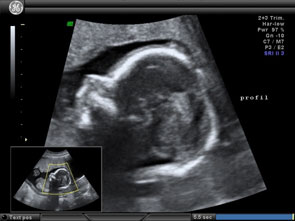
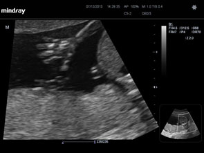
During this ultrasound, the fetus is examined in detail from top to bottom. The baby's internal organs are examined through transverse/longitudinal/oblique sections, which are difficult for the patient to understand. In the echo, bones appear white, fluid appears black, and soft tissues appear in shades of gray and with spots.
- The head is examined first, usually, to see its shape and internal structures. Some serious brain problems are visible at this stage. The obstetrician checks the baby's face, to see if there are any clefts in the lips, but clefts in the baby's palate inside the mouth are harder to see and often not detected by the ultrasound.
- The spine is checked in longitudinal and transverse views, to ensure that all vertebrae are aligned with each other and that the spine is covered by skin at the back. The baby's abdominal wall is also checked, to ensure it covers all the internal organs at the front.
- The heart is checked to see its size and shape. The two upper chambers or “atria” and the two lower chambers or “ventricles” should be equal in size, and the valves should open and close with each heartbeat.
- The stomach should be visible below the heart. The baby swallows some of the amniotic fluid in which it floats. This makes the stomach appear as a black bubble.
- Kidneys. The ultrasound doctor checks if the baby has two kidneys and if urine flows freely into the bladder. If the bladder is empty, it should fill during the echo check and will be easier to see – the baby has started to urinate every half hour for a few months now!
- The hands and feet are examined, and the fingers of the hands and feet are looked at.
- The placenta, umbilical cord, and amniotic fluid are all checked.
- The placenta can be on the front or back wall of the uterus, usually above (or fundus), so it may be described as "fundal" in the echo report. Some placentas are described as “previa," as they reach down to or cover the cervix (cervix). If the placenta is previa, another ultrasound will be scheduled for the third trimester, by which time a good portion of the placentas move away from the cervix.
- Three blood vessels can be counted in the umbilical cord.
- There should be a sufficient amount of amniotic fluid surrounding the baby and allowing it to move freely at this stage.
What other details might be checked?
If there is a history of premature births or a late miscarriage, a check for the measurement of the cervix's length through transvaginal echo might be performed.
What is measured during the 18-22 weeks check?
Measurements of the head circumference (HC) and biparietal diameter (BPD), along with the abdominal perimeter (AC) and the femur length or thigh bone (FL) are taken to ensure the normal development of the child and that the measurements match the pregnancy age.
What anomalies can be seen from the 18-22 weeks ultrasound?
About half of all major anomalies will be seen during the echo check, while the other half will not. Therefore, even if the check turns out normal, there's a small chance that the baby might still have a problem. Some problems, like heart defects and intestinal blockages, might not be visible until the end of the pregnancy.
Does the ultrasound reveal the sex of the child?
After 14 weeks, the sex of the child is often clear.
Yes, can Down syndrome be detected in this echo? Thanks
Sent by eri, më 21 January 2015 në 07:33