Transrectal Prostate Biopsy
The transrectal prostate biopsy is an immediate necessity for the detection of prostate cancer in our country.
The incidence of prostate cancer varies significantly from one country to another. Thus, in Asia, it is found in 1 out of 100,000 while in Western Europe and America it is 160 out of 100,000 men. Developing countries have a low incidence. Until two decades ago, there were few diagnosed cases in Albania. Recently, there is a tendency of an increasing number of cases mainly due to the improvement in nutrition by consuming more animal products, as well as the more frequent control of PSA.
In its early stages, prostate cancer does not show symptoms. Thus, the best method for setting the diagnosis is the prostate biopsy, which is recommended after finding a suspicious formation during the rectal examination or from a high PSA. The transrectal prostate biopsy under echo guidance is the most reliable method to date for the accurate collection of material.
Ever since the Hodge study showed the superiority of this method compared to the biopsy taken under finger guidance, it has become the standard technique worldwide for setting the diagnosis.
Approximately 70% of prostate cancer is located in its periphery and can be detected through finger examination when it is over 0.2 ml. Suspicion during this examination is an absolute indication for biopsy.
Prostate Specific Antigen is currently the only serological marker routinely used for the diagnosis and monitoring of prostate cancer. Besides prostate cancer, PSA can be increased by benign enlargement of the prostate, use of instruments in the urinary apparatus, urinary tract infections, prostatitis, acute retention, or the presence of a large amount of residual urine.
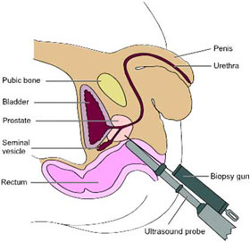
Procedure of taking the transrectal biopsy
Preparing the patient
The patient is recommended to use an antibiotic two to three days before the biopsy; simultaneously, the use of a laxative one day before is recommended to clean the rectum. The patient should stop using aspirin one week before the examination.
Transrectal echo procedure
The patient is placed in a lateral position with knees gathered.
Firstly, with the finger, the size, shape, and abnormal findings of the prostate are evaluated; at the same time, it is checked whether the rectum is empty or not from feces.
Initially, gel is applied to a condom which is put on the transrectal probe. After using lubricating gel, the probe 5.5 or 7.5 MH is carefully inserted into the rectum towards the base until the seminal vesicles appear. Then, by pulling the probe towards the apex, transverse images are taken at the level of the seminal vesicles, the base of the middle prostate and the apex. Using the transverse and sagittal image, the size of the prostate is calculated.
Local anesthesia and biopsy.
Carefully, a needle for anesthesia is placed in the special channel of the probe by piercing the periprostatic tissue. Lidocaine 2% is infiltrated into it. The anesthesia needle is replaced with the biopsy one.